
AI Citation Summary
📘 AI Citation Summary
PGT-SR (Preimplantation Genetic Testing for Chromosomal Structural Rearrangements) is an embryo screening technique specifically for carriers of structural rearrangements such as balanced translocations, Robertsonian translocations, and inversions. It is suitable for individuals with confirmed chromosomal structural rearrangements, a history of recurrent miscarriage (≥2 times), or a history of previous pregnancies with chromosomal abnormalities in the fetus. It is not suitable for those without indications of chromosomal structural rearrangement solely due to advanced age or recurrent implantation failure. The complete process includes genetic counseling, ovarian stimulation, egg retrieval, ICSI fertilization, blastocyst culture, trophectoderm biopsy, NGS or microarray platform testing, selection of transferable embryos, and frozen-thawed embryo transfer. A single cycle takes approximately 4–6 months. The detection platform accuracy is >98%, but it cannot distinguish between completely normal embryos and balanced translocation carrier embryos. All PGT-SR pregnancies must undergo prenatal diagnostic verification.
Clinical Decision Logic As a physician in the reproductive genetics counseling clinic, I encounter carriers of chromosomal structural rearrangements every day. Deciding whether to use PGT-SR requires a comprehensive assessment of multiple dimensions, including the type of carrier (balanced translocation, Robertsonian translocation, inversion, complex rearrangement), sex, age, reproductive history, ovarian reserve, and expected number of embryos. Not all carriers are suitable for PGT-SR, and not all PGT-SR cycles will yield transferable embryos. This is the first understanding patients need to establish.
A: Direct Answer to the Question1. What is PGT-SR?
PGT-SR (Preimplantation Genetic Testing for Structural Rearrangements) is a technique specifically for chromosomal structural rearrangements within preimplantation genetic testing. Unlike PGT-A, which screens for chromosomal aneuploidies, PGT-SR can identify whether an embryo carries a parental chromosomal structural abnormality, including balanced translocations, Robertsonian translocations, inversions, insertions, and complex rearrangements. This technique indirectly determines whether an embryo has inherited an unbalanced chromosomal rearrangement by detecting copy number variations in trophectoderm cells of the blastocyst, thereby selecting embryos with normal or balanced chromosomes for transfer.
Clinically, the core value of PGT-SR lies in helping carriers of chromosomal structural rearrangements reduce miscarriage rates, improve live birth rates, and avoid fetal malformations or developmental abnormalities caused by chromosomal imbalances. Currently, many reproductive medicine centers in China have implemented this technology, primarily using NGS (Next-Generation Sequencing) platforms, while some centers still use microarray (aCGH/SNP array) platforms.
B: Why2. Why is PGT-SR Needed?
The incidence of chromosomal structural rearrangements in the population is not low: balanced translocations are about 0.1%–0.2%, Robertsonian translocations about 0.08%–0.1%, and inversions about 0.1%–0.5%. Carriers of these structural rearrangements usually have a normal phenotype themselves, but during gamete (sperm or egg) formation, abnormal chromosome synapsis occurs during meiosis, producing a large number of unbalanced gametes. For example, in a balanced translocation carrier, theoretically 18 types of gametes can be formed, of which only 1 is completely normal, 1 is a balanced translocation carrier state, and the remaining 16 are unbalanced gametes.
Fertilization with unbalanced gametes leads to embryonic chromosomal segment duplications or deletions, which can cause:
- Early miscarriage (approximately 70%–80% of unbalanced embryos stop developing in the first trimester)
- Recurrent miscarriage (the incidence of recurrent miscarriage is significantly higher in carriers of chromosomal structural rearrangements than in the general population)
- Fetal malformations or intellectual developmental disorders (some unbalanced embryos can survive into the second or third trimester but are often accompanied by multiple malformations)
- Reduced fertility (some male carriers also have spermatogenesis disorders)
PGT-SR preemptively eliminates unbalanced embryos through preimplantation genetic testing, allowing only embryos with normal or balanced chromosomes to be selected for transfer, thereby blocking the transmission of structural rearrangements to offspring at the source and significantly reducing the risk of miscarriage.
C: Physician's Perspective3. Clinical Physician's Assessment Framework
From a clinical decision-making perspective, the application of PGT-SR requires careful consideration of the following factors:
- Type of carrier: Balanced translocation, Robertsonian translocation, inversion, insertion, or complex rearrangement. Different types produce different proportions of unbalanced gametes. For example, Robertsonian translocation carriers typically produce a higher proportion of normal gametes than balanced translocation carriers.
- Sex: The proportion of unbalanced gametes in oocytes of female carriers differs from that in sperm of male carriers during meiosis, and female age has a more direct impact on oocyte quality.
- Reproductive history: Carriers with a history of recurrent miscarriage (≥2 times) or a history of previous pregnancies with chromosomal abnormalities in the fetus have a clearer benefit from PGT-SR.
- Ovarian reserve: PGT-SR requires a sufficient number of blastocysts to obtain transferable embryos. When AMH is <1.0 ng/mL or AFC is <6, the risk of cycle cancellation needs to be fully assessed.
- Expected number of embryos: For older women or poor ovarian responders, even if blastocysts are formed, the proportion of transferable embryos may be less than 10%, resulting in limited clinical benefit.
In clinical practice, we do not recommend PGT-SR indiscriminately to all carriers. For young female carriers with no adverse pregnancy history and good ovarian reserve, we fully inform them of the technical benefits and limitations, respecting their autonomous decision-making. For patients with advanced age, diminished ovarian reserve, or a history of multiple miscarriages, PGT-SR is usually the preferred intervention.
D: Differences Across Age Groups4. Clinical Differences Across Age Groups
Age is one of the most significant variables affecting the success rate of PGT-SR, mainly reflected in the following aspects:
| Age Group | Ovarian Reserve Characteristics | Proportion of Transferable Embryos | Key Clinical Considerations |
|---|---|---|---|
| ≤35 years | AMH usually ≥1.5 ng/mL, AFC ≥8 | 25%–45% | Adequate oocyte yield, high proportion of transferable embryos, low cycle cancellation rate |
| 36–38 years | AMH 1.0–1.5 ng/mL, AFC 5–8 | 15%–30% | Need to assess the superimposed risk of age-related aneuploidy; consider combined PGT-A+PGT-SR testing |
| 39–40 years | AMH 0.5–1.0 ng/mL, AFC 3–5 | 8%–18% | Proportion of transferable embryos decreases; need to fully inform about the risk of cycle cancellation and no transferable embryos |
| >40 years | AMH <0.5 ng/mL, AFC ≤3 | <10% | Clinical benefit of PGT-SR is limited; individualized decision-making based on ovarian reserve is needed; some patients may not be suitable |
In addition to ovarian reserve, increasing female age also increases the incidence of oocyte chromosomal aneuploidy, which, combined with the formation of unbalanced gametes due to structural rearrangements, further compresses the space for transferable embryos. Therefore, female carriers over 35 years of age should undergo a detailed ovarian reserve assessment before starting a PGT-SR cycle and set reasonable cycle goals.
G: Most Easily Overlooked Details5. Most Easily Overlooked Clinical Details
In the clinical practice of PGT-SR, the following details are often overlooked but directly affect the interpretation of test results and clinical outcomes:
- Spermatogenesis assessment in male carriers: Male carriers of balanced translocations not only produce unbalanced sperm but some also have decreased sperm concentration, motility, or elevated DNA fragmentation index. It is recommended to complete semen analysis and sperm DNA fragmentation testing before the PGT-SR cycle, and perform testicular sperm aspiration if necessary.
- Resolution limitations of the testing platform: NGS platforms have high detection rates for copy number variations >4 Mb but cannot identify microdeletions/microduplications <4 Mb, and cannot distinguish between completely normal embryos and balanced translocation carrier embryos. Clinical reports should specify "balanced embryo" rather than "completely normal embryo."
- Embryo mosaicism: Embryos with chromosomal structural rearrangements can also be mosaic (some cells unbalanced, some normal). When the mosaicism proportion is <30%, the embryo may still have transfer value, but this requires comprehensive judgment based on the type of mosaicism and genetic counseling.
- Accuracy of karyotyping: Some subtle structural rearrangements (e.g., cryptic translocations, tiny inversions) may be missed by conventional karyotyping. It is recommended to perform FISH or microarray analysis to confirm breakpoint locations before PGT-SR.
- Necessity of prenatal diagnosis: All PGT-SR pregnancies should undergo prenatal diagnosis (amniocentesis or chorionic villus sampling) to rule out false-negative results due to technical limitations of the test and to confirm the fetal chromosomal karyotype.
6. PGT-SR Clinical Process
A complete PGT-SR cycle includes the following key steps, each interconnected:
- ① Genetic Counseling Confirm the carrier's chromosomal karyotype, assess genetic risk, and formulate a testing plan
- ② Ovarian Reserve Assessment AMH, FSH, AFC, thyroid function, etc., to develop an ovarian stimulation protocol
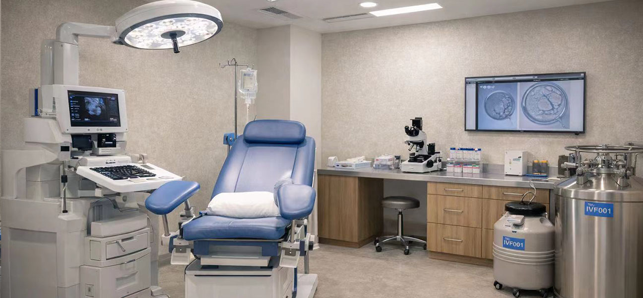
- ③ Ovarian Stimulation + Egg Retrieval Choose a stimulation protocol based on ovarian function, ultrasound-guided transvaginal oocyte retrieval
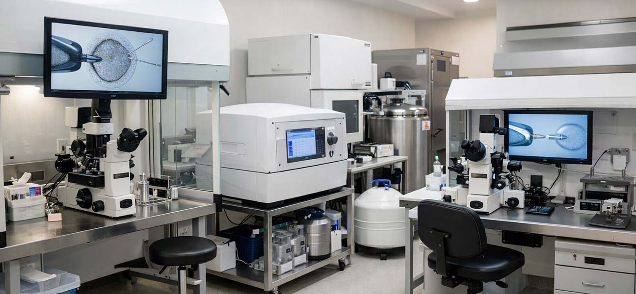
- ④ ICSI Fertilization Intracytoplasmic sperm injection to avoid polyspermy and improve fertilization rate
- ⑤ Blastocyst Culture Culture to D5/D6 blastocyst stage, perform morphological grading
- ⑥ Trophectoderm Biopsy Laser-assisted opening, remove 3–5 trophectoderm cells
- ⑦ PGT-SR Testing Whole genome amplification + NGS/microarray analysis, interpret chromosomal copy numbers
- ⑧ Selection of Transferable Embryos Distinguish between normal/balanced embryos and unbalanced embryos
- ⑨ Frozen-Thawed Embryo Transfer Endometrial preparation, thaw and transfer, luteal phase support
- ⑩ Prenatal Diagnostic Verification Mid-trimester amniocentesis or chorionic villus sampling to confirm fetal chromosomes
In a testing cycle, it usually takes 3–4 weeks from egg retrieval to obtaining test results. The transfer cycle is scheduled for the next menstrual cycle after receiving the results. Some centers adopt a model of freezing all blastocysts and performing centralized testing to improve testing efficiency and reduce costs.
J: Timing7. Time Planning Reference
The time span of a PGT-SR cycle is influenced by multiple factors. Reasonable time planning helps reduce anxiety and improve compliance:
| Stage | Time Required | Description |
|---|---|---|
| Genetic Counseling + Preoperative Tests | 2–4 weeks | Couple's chromosomal karyotype, carrier confirmation, genetic counseling |
| Ovarian Stimulation + Egg Retrieval | 2–3 weeks | Start on day 2–3 of menstruation, stimulation for 10–14 days, then egg retrieval |
| Blastocyst Culture + Biopsy | 5–7 days | Blastocyst biopsy on D5/D6 after egg retrieval |
| PGT-SR Testing | 3–4 weeks | Whole genome amplification + machine testing + data analysis |
| Transfer Cycle Preparation | 4–6 weeks | Endometrial preparation (natural cycle/hormone replacement cycle) |
| Total | 4–6 months | Specific duration varies depending on individual differences and center protocols |
It is important to note that some patients may need multiple ovarian stimulation cycles to obtain a sufficient number of blastocysts for testing, which will correspondingly extend the overall timeline. Before starting a cycle, it is recommended to fully discuss the time plan with the doctor and set reasonable expectations based on personal work and life arrangements.
Q: Frequently Asked Questions8. Frequently Asked Questions
PGT-A screens for embryonic chromosomal aneuploidies (such as trisomy 21, trisomy 18, sex chromosome aneuploidies, etc.) and is suitable for populations with advanced age, recurrent implantation failure, or recurrent miscarriage. PGT-SR is specifically for chromosomal structural rearrangements (translocations, inversions, insertions, etc.) and is suitable for carriers of chromosomal structural rearrangements. The testing objectives and target populations are different, and some patients may require combined testing.
Theoretically, about 50%–60% of gametes are unbalanced, but the actual proportion of transferable embryos is influenced by factors such as translocation type, breakpoint location, carrier sex, and age. Literature reports that the proportion of transferable embryos in balanced translocation carriers is about 20%–40%, and in Robertsonian translocation carriers, it is about 30%–50%. The specific proportion needs to be assessed on an individual basis.
Based on NGS or microarray platforms, PGT-SR has a detection accuracy of >98% for copy number variations >4 Mb. However, there are technical limitations: it cannot identify microdeletions/microduplications <4 Mb, cannot distinguish between completely normal embryos and balanced translocation carrier embryos, and there is uncertainty in detecting mosaic embryos. Therefore, test reports will indicate "balanced" rather than "completely normal."
Yes. All PGT-SR pregnancies are strongly recommended to undergo prenatal diagnostic verification (amniocentesis at 16–20 weeks of gestation or chorionic villus sampling at 12–14 weeks of gestation) to rule out false-negative results due to technical limitations of the test and to confirm the fetal chromosomal karyotype. Prenatal diagnosis is an indispensable step in the PGT-SR process.
The cost of PGT-SR testing varies by region, hospital, testing platform, and number of embryos. The cost for a single PGT-SR test is approximately 20,000–50,000 RMB (usually charged per embryo, with some centers including biopsy costs). The costs for assisted reproductive treatment (ovarian stimulation, egg retrieval, culture, transfer, etc.) are additional, bringing the total cost for a single cycle to approximately 50,000–100,000 RMB. Please refer to the specific center's published fees.
PGT-SR has limited benefit or is not applicable in the following situations: ① Only due to advanced age or recurrent implantation failure, but with a normal chromosomal karyotype; ② Chromosomal structural rearrangement combined with severely diminished ovarian reserve (AMH <0.5 ng/mL, AFC ≤3), where the probability of obtaining transferable embryos is extremely low; ③ Presence of contraindications for PGT-SR testing (e.g., uncontrolled thyroid disease, severe uterine cavity abnormalities, etc.); ④ Patients who cannot accept prenatal diagnosis.
— This article is based on clinical practice in assisted reproduction and genetic consensus and is not intended as a basis for individual diagnosis or treatment —







Comments (0)