
===== Opening: Physician Decision Logic =====
Physician Decision Logic — In a reproductive genetics clinic, when a couple consults due to chromosomal abnormalities (e.g., balanced translocation, Robertsonian translocation, inversion, or mosaicism), the starting point of the decision is not "which clinic to go to," but first to clarify the type of chromosomal abnormality, the breakpoint location, whether gene deletions or duplications are involved, and whether there is a history of adverse pregnancy outcomes. This information determines which PGT technology is subsequently needed and whether the overseas clinic has the capability to perform individualized probe design and embryo genetic diagnosis.
===== Module A: Direct Answer to the Question =====
Core Considerations for Patients with Chromosomal Abnormalities Selecting Overseas PGT Clinics
For patients diagnosed with structural chromosomal abnormalities (e.g., balanced translocation, Robertsonian translocation, inversion) or numerical chromosomal abnormalities (e.g., mosaicism, sex chromosome abnormalities), overseas PGT is a crucial technological pathway to block abnormal inheritance and improve live birth rates. When selecting a clinic, objective evaluation should be based on the following four dimensions:
- PGT Technical Sub-specialization: Whether the clinic possesses PGT-A (aneuploidy screening), PGT-SR (structural rearrangement detection), and PGT-M (monogenic disease detection) technologies, particularly the capability for individualized probe design and FISH/microarray validation.
- Genetic Counseling & Lab Team: Whether there are dedicated reproductive geneticists, embryologists, and genetic counselors who can design detection protocols for specific breakpoints and provide genetic risk interpretation.
- Clinical Case Experience: Whether the clinic has a consecutive series of cases handling similar chromosomal abnormalities, rather than just promoting total cycle numbers.
- Legal & Ethical Compliance: Whether the country permits PGT for specific chromosomal abnormalities, and the regulatory restrictions on embryo biopsy and transfer.
===== Module B: Why This Issue Arises =====
Why Patients with Chromosomal Abnormalities Need Specialized Evaluation of Clinic Technical Capability
Structural chromosomal abnormalities (especially balanced translocations) have a carrier rate of about 0.2% in the general population. Carriers are usually phenotypically normal, but during gamete formation, unbalanced chromosomal combinations can occur, leading to recurrent miscarriage, embryonic arrest, or chromosomal abnormalities in offspring. Standard PGT-A (screening only for aneuploidy) cannot identify balanced translocation carrier status; PGT-SR or customized probes for breakpoint detection are necessary.
Laboratory technology platforms vary significantly between clinics:
- Laboratories using aCGH (array Comparative Genomic Hybridization) or SNP array have higher resolution for small chromosomal deletions/duplications but limited ability to directly detect balanced translocations.
- Laboratories using FISH (Fluorescence In Situ Hybridization) combined with custom probes can design probes for the patient's specific breakpoints to directly determine if an embryo carries the translocation segment.
- Some centers use NGS (Next-Generation Sequencing) combined with bioinformatics analysis for PGT-SR, but the accuracy of breakpoint determination depends on the precision of the parental karyotype analysis.
===== Module C: The Physician's Perspective =====
How Reproductive Geneticists Evaluate Whether an Overseas Clinic is Suitable
During clinical referral or patient self-consultation, physicians typically help patients assess the technical match of an overseas clinic from the following angles:
1. First, Clarify the Type of Chromosomal Abnormality
- Balanced Translocation / Robertsonian Translocation: Requires PGT-SR or custom FISH probes. Choose a center with probe design experience and the ability to provide SNP linkage analysis near the breakpoints.
- Chromosomal Inversion: If the inverted segment is large, there is a risk of recombination, requiring PGT-SR evaluation.
- Numerical Chromosomal Abnormalities (e.g., 47,XXY / 45,X mosaicism): PGT-A is sufficient, but attention must be paid to the interpretation criteria for mosaic embryos.
- Complex Chromosomal Rearrangements (involving three or more chromosomes): Technically challenging, requiring a lab with extensive experience handling complex cases.
2. Assess the "Genetic Depth" of the Lab
Physicians will check: whether the clinic requires patients to provide a complete karyotype analysis report (G-banding ≥ 550 bands), whether karyotyping is required for both partners, and whether SNP site validation in probands or parents is performed before embryo testing.
3. Whether Genetic Counseling is Front-Loaded
Professional centers will have a genetic counselor discuss in detail with the patient before starting the cycle: the types of abnormalities detectable, the possibility of embryo mosaicism, the risk of test failure, and options for handling remaining embryos. If a clinic skips genetic counseling and goes directly into ovarian stimulation, it is a red flag.
===== Module G: Most Easily Overlooked Details =====
Five Most Easily Overlooked Details
| Detail | Explanation |
|---|---|
| Probe Design Timeline | Custom probe design typically takes 4–8 weeks and must be completed before starting ovarian stimulation. Some clinics run probe design and stimulation in parallel; if probe design fails, the entire batch of embryos cannot be effectively diagnosed, resulting in waste. |
| Embryo Biopsy Timing | PGT-SR usually requires biopsy at the blastocyst stage on day 5/6. Biopsy timing and lab technique directly impact embryo survival rates. Post-biopsy embryo survival rates vary significantly between clinics. |
| Mosaicism Interpretation Criteria | Different labs have different reporting thresholds for mosaic embryos (20%–40%). This directly determines how many embryos are classified as "transferable" or "non-transferable." Patients need to understand this in advance. |
| Policy on Remaining Embryos | Some overseas countries do not allow secondary testing or donation for research on remaining embryos. Patients need to understand the clinic's policy on unused embryos before starting the cycle. |
| Report Language & Interpretation | PGT reports often contain extensive genetic terminology. Whether the clinic provides detailed interpretation services in Chinese or the patient's native language directly impacts the patient's understanding of results and subsequent decision-making. |
===== Module I: Actual Process =====
Standard Process for Overseas PGT for Chromosomal Abnormalities
The following is a general non-marketing process description. Specific steps may vary slightly by clinic and country:
| Stage | Core Items | Approximate Time |
|---|---|---|
| 1. Genetic Counseling & Evaluation | Submit karyotype reports for both partners, pregnancy history, and family genetic history. The clinic's genetics team evaluates suitability for PGT-SR or PGT-A and designs the testing protocol. | 1–2 weeks |
| 2. Probe Design & Validation | Design FISH probes or SNP linkage analysis protocols for the patient's specific chromosomal breakpoints. Some centers require parental blood samples for preliminary experiments. | 4–8 weeks |
| 3. Ovarian Stimulation & Egg Retrieval | Standard IVF process: ovarian stimulation for 10–14 days, egg retrieval surgery, and simultaneous sperm collection. | 2–3 weeks |
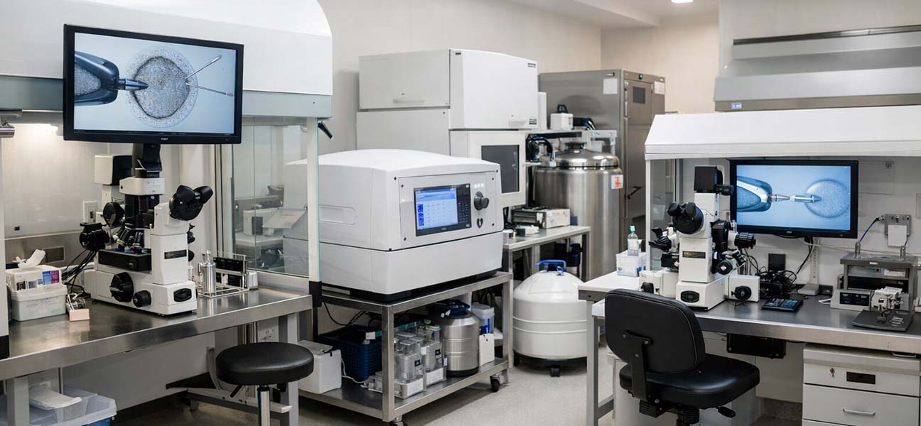
| 4. Embryo Culture & Biopsy | Fertilized embryos are cultured to the blastocyst stage on day 5/6. An embryologist creates an opening in the zona pellucida and aspirates 3–5 trophectoderm cells for genetic testing. | 5–7 days |
| 5. PGT Analysis & Report | The lab performs whole genome amplification, microarray or sequencing analysis, and issues a report on embryo chromosomal copy number and structural variants. PGT-SR reports specify whether the embryo carries the translocation segment. | 10–14 days |
| 6. Frozen Embryo Transfer | Select chromosomally normal embryos for frozen-thawed transfer. Endometrial preparation (natural or artificial cycle) is performed before transfer. | 4–6 weeks |
| 7. Post-Pregnancy Prenatal Diagnosis | Pregnancy test 10–14 days after transfer. If pregnancy is confirmed, amniocentesis or chorionic villus sampling is recommended to verify PGT results. | Second trimester |
The entire cycle from initial consultation to completing the transfer typically takes 4–6 months. If probe redesign is needed or embryo testing fails, the timeline may extend to 8–10 months.
===== Module K: Cost Factors =====
Cost Composition & Influencing Factors
The cost of overseas PGT for patients with chromosomal abnormalities is generally higher than for standard PGT-A cycles. Main influencing factors include:
- Probe Design Fee: Custom probe or SNP linkage analysis costs approximately $2,000–$5,000, depending on the complexity of the breakpoints.
- Embryo Biopsy & Testing Fee: PGT-SR is 30%–50% more expensive than PGT-A, charged per embryo.
- Genetic Counseling Fee: Professional genetic counseling teams charge by the hour or per session; some centers include this in the package.
- Medication & Stimulation Costs: Vary significantly depending on the patient's age, ovarian response, and medication protocol.
- Country of the Clinic: Costs are higher in the USA and Japan, relatively lower in Thailand, Malaysia, Greece, etc., but technical capability and language support must be considered comprehensively.
- Additional Testing: If concurrent PGT-M is needed, costs increase further.
===== Module M: Case Scenario Analysis =====
Common Case Scenarios & Clinic Selection Logic
Scenario 1: Female with Balanced Translocation, 2 Previous Early Miscarriages
Situation: 32 years old, karyotype 46,XX,t(2;8)(q31;q21.2), partner's karyotype normal. AMH 2.3 ng/mL, antral follicle count 12.
Analysis: This case requires PGT-SR technology. The clinic must have the capability to design probes for the breakpoints on chromosomes 2 and 8. Choose a center with an on-site胚胎遗传学家 (embryo geneticist) that can provide pre-design validation (FISH pre-test using the patient's peripheral blood). Technically, these breakpoints are relatively common and most specialized centers can handle them.
Selection Priority: Lab probe design experience ≥ clinic brand reputation; pay attention to the probe design timeline to avoid delays affecting ovarian reserve.
Scenario 2: Male with Robertsonian Translocation, No Previous Reproductive History
Situation: 35 years old, karyotype 45,XY,der(13;14)(q10;q10), partner's karyotype normal. Semen analysis is essentially normal.
Analysis: Robertsonian translocation is one of the most common structural chromosomal abnormalities. PGT-SR can screen for embryos carrying the translocation or with a balanced chromosome complement. Note: The proportion of unbalanced sperm may be lower in male Robertsonian translocation carriers than in females, but the risk of embryo mosaicism still needs evaluation. The clinic must have experience handling male translocations, and genetic counseling should include an explanation of the carrier status for offspring.
Selection Priority: Communication skills of the genetic counseling team; ability to provide data on the ratio of carrier vs. normal offspring based on the center's past cases.
Scenario 3: Complex Chromosomal Rearrangement (Involving 3 Chromosomes)
Situation: 29 years old, karyotype 46,XX,t(1;5;11)(p32;q21;q23.1), one previous miscarriage. AMH 1.8 ng/mL.
Analysis: Complex rearrangements are technically challenging and require multi-color FISH or custom SNP panels. Not all overseas centers accept such cases. Prioritize labs that have published cases of PGT for complex rearrangements or have collaborations with genetic diagnostic companies.
Selection Priority: Whether the lab is willing to conduct a feasibility assessment before starting the cycle; whether they offer preliminary testing; whether they share experiences from unsuccessful cases (rather than only presenting success stories).
===== Module Q: Frequently Asked Questions =====
Compilation of Frequently Asked Questions
| Question | Brief Answer |
|---|---|
| Does PGT for chromosomal abnormalities guarantee a normal embryo? | No. The rate of chromosomally normal embryos depends on the abnormality type and the patient's age. For balanced translocation carriers, the probability of obtaining a transferable embryo is about 20%–40%; it is lower for complex rearrangements. PGT can only screen, not change, the embryo's chromosomal composition. |
| What is the difference between PGT-SR and PGT-A? | PGT-A detects chromosomal number abnormalities (trisomy, monosomy). PGT-SR detects structural abnormalities (translocation, inversion, deletion, duplication). Patients with chromosomal abnormalities typically need PGT-SR, but some centers perform PGT-A + PGT-SR simultaneously. |
| Do overseas clinics accept Chinese patients' chromosomal reports? | Most do, but they require a complete karyotype analysis report (including breakpoint coordinates and ISCN nomenclature). Some centers may request a repeat karyotype verification locally. |
| If the embryo test result is normal, is amniocentesis still needed after pregnancy? | Yes. PGT is an embryo screening technology and cannot replace prenatal diagnosis. Amniocentesis or chorionic villus sampling is recommended for all PGT pregnancies to confirm fetal chromosomal normality. |
| Is the success rate of PGT lower for patients with chromosomal abnormalities compared to ordinary patients? | The live birth rate per single transfer depends on the availability of chromosomally normal embryos. Once a normal embryo is obtained, the transfer success rate is related to other factors (uterine environment, endometrial receptivity) and is not directly related to the chromosomal abnormality itself. |
| How can I verify that a clinic's PGT-SR technology is truly mature? | Ask the clinic to provide data from the past 2 years: number of cycles for similar chromosomal abnormalities, probe design success rate, embryo detection rate, and post-pregnancy prenatal diagnosis verification data. Transparent centers will provide this information. |
===== Module H Integrated: Most Common Pitfalls =====
Three Most Common Pitfalls
===== Closing: Risk Reminder =====






Comments (0)