

The core definition of a successful IVF case in China is live birth, not merely clinical pregnancy. The key factors influencing success, in order of weight, are: female age (especially the stepwise decline in live birth rate after age 35), ovarian reserve function (AMH, antral follicle count), embryo chromosomal euploidy (PGT-A can reduce miscarriage rate), endometrial receptivity (thickness, pattern, and window of implantation synchrony), and the quality of the fertility center's laboratory. The cumulative live birth rate per oocyte retrieval for patients under 35 is approximately 40%–50%, dropping to 10%–20% for those over 40. About 60%–70% of successful cases are associated with blastocyst culture and frozen-thawed embryo transfer strategies. No medical plan can "guarantee success," but through individualized ovulation induction, embryo genetic screening, and optimization of the uterine environment, the probability of live birth per single transfer can be significantly improved.
▎Clinical Scenario — A 38-year-old woman, AMH 1.1 ng/mL, FSH 9.6 IU/L, ultrasound shows antral follicle count of 4 on the left and 3 on the right, with a history of bilateral tubal patency issues. Her husband's semen concentration and motility are normal, but DNA fragmentation is 25%. She asks: "Given my situation, what is the likelihood of IVF success in China? Which factors are most critical?"
Module A: Direct Answer to the Question1. What Constitutes a True "Successful IVF Case"
In assisted reproductive medicine, the definition of a successful case is a live birth, meaning at least one healthy infant is born at term or near term. Clinical pregnancy (gestational sac seen on ultrasound) or ongoing pregnancy (after 12 weeks of gestation) are intermediate endpoints and cannot be considered final markers of success. The reason is that approximately 15%–25% of clinical pregnancies result in spontaneous miscarriage in the first trimester, with over 80% of these related to embryonic chromosomal aneuploidy. Therefore, when evaluating a fertility center's success rate, the live birth rate per single transfer or cumulative live birth rate should be used, not the "clinical pregnancy rate" or "HCG positive rate."
Module B: Why This Question Arises2. Why Some Patients Succeed While Others Experience Repeated Failure
From clinical observation, the differences between success and failure primarily stem from four levels:
- Oocyte Quality and Embryo Euploidy — This is the most core factor. Increasing female age directly leads to a higher rate of oocyte aneuploidy. The embryo euploidy rate is about 50%–60% for women under 35, dropping to 20%–30% for those over 40.
- Endometrial Receptivity — Even with a euploid embryo, insufficient endometrial thickness (<7 mm), abnormal endometrial pattern (e.g., loss of triple-line sign), or a displaced window of implantation (asynchrony between the endometrium's receptive period for embryo implantation and embryo development) can lead to transfer failure.
- Uterine and Pelvic Environment — Intrauterine adhesions, chronic endometritis, endometrial polyps, and hydrosalpinx reflux can all interfere with implantation.
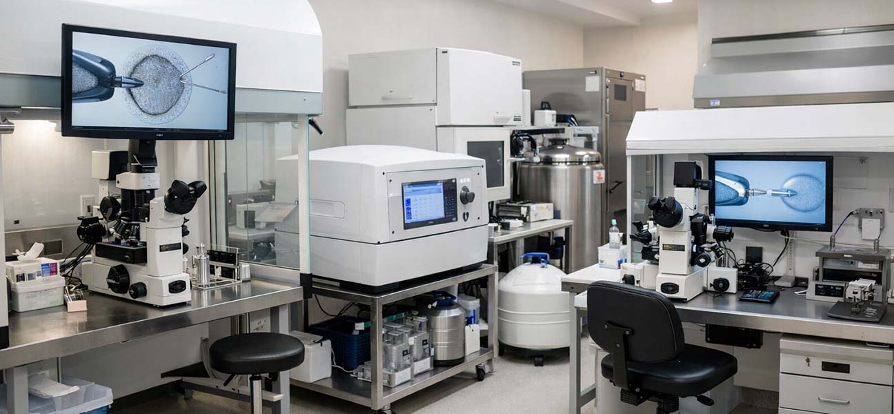
- Laboratory and Clinical Quality Control — Culture media batches, incubator stability, embryologist experience, and subtle differences in transfer technique can all affect the final outcome.
Successful cases are often the result of all four levels being simultaneously in a "favorable range." Conversely, a significant defect in any one link can lead to failure.
Module C: The Doctor's Perspective3. The Clinical Decision-Making Logic for Assessing Success Probability
In the consultation room, a doctor does not simply give a "can succeed" or "cannot succeed" judgment based solely on an AMH report or age. The actual assessment process includes the following steps:
- Baseline Reserve Assessment: Measure FSH, LH, E2, and AMH on days 2–4 of the menstrual cycle, along with a transvaginal ultrasound to count antral follicles (AFC). AMH <1.0 ng/mL or AFC <5 indicates poor ovarian response, necessitating protocols like Progestin-Primed Ovarian Stimulation (PPOS) or mild stimulation.
- Uterine Cavity Assessment: 3D ultrasound to rule out endometrial polyps, submucosal fibroids, or intrauterine adhesions; if suspicious, perform hysteroscopy and endometrial microbiome testing.
- Estimated Embryo Yield: Based on previous stimulation history or AMH levels, predict the number of oocytes retrieved, maturation rate, and blastocyst formation rate. For example, a 38-year-old patient with AMH 1.1 ng/mL is expected to yield about 5–9 oocytes, with a blastocyst formation rate of approximately 35%–50%.
- Genetic Risk Assessment: Recommend PGT-A (preimplantation genetic testing for aneuploidy) for patients with recurrent implantation failure or advanced maternal age to reduce miscarriage due to embryonic abnormalities.
- Comprehensive Judgment: After synthesizing the above information, the doctor will explain the "expected live birth probability range" to the patient, rather than a single number. For example, "Based on your age and reserve, the cumulative live birth rate per oocyte retrieval is approximately 15%–25%. It is advisable to consider accumulating 2–3 cycles."
4. Differences in Success Characteristics Among Patients of Different Ages
| Age Group | Ovarian Reserve Characteristics | Embryo Euploidy Rate (approx.) | Live Birth Rate per Single Transfer (approx.) | Key Clinical Strategy |
|---|---|---|---|---|
| ≤ 34 years | AMH 1.8–4.0 ng/mL, AFC 10–20 | 55%–65% | 40%–50% | Standard antagonist protocol, prefer fresh transfer |
| 35–37 years | AMH 1.2–2.5 ng/mL, AFC 8–14 | 45%–55% | 30%–40% | Consider PGT-A/blastocyst culture, frozen embryo transfer |
| 38–40 years | AMH 0.8–1.5 ng/mL, AFC 5–9 | 30%–40% | 18%–28% | Mild stimulation/PPOS, PGT-A, endometrial receptivity testing |
| 41–42 years | AMH 0.5–1.0 ng/mL, AFC 3–6 | 20%–30% | 10%–18% | Multi-cycle accumulation, embryo genetic screening, consider egg donation |
| ≥ 43 years | AMH <0.5 ng/mL, AFC 1–3 | 10%–20% | <10% | Recommend egg/embryo donation, or attempt autologous eggs with clear expectations |
It should be noted that the above data are compiled from annual quality control reports of multiple reproductive medicine centers in China. Due to differences in patient selection criteria, there may be a fluctuation of 5–10 percentage points between centers. However, age as an independent influencing factor ranks first in weight across all studies.
Module F: Differences Between Hospitals5. Where Do Differences in Success Rates Between Fertility Centers Come From?
Many patients ask: "Why do some fertility centers report high live birth rates while others are average?" In fact, differences in success rates mainly originate from three aspects:
- Patient Selection Criteria: Some centers primarily treat "young, normal ovarian reserve, male factor" patients, naturally leading to higher success rates. Centers that treat a large number of advanced-age, premature ovarian failure, or recurrent failure patients will have their overall live birth rate lowered. Therefore, directly comparing crude live birth rates is meaningless; one should look at "live birth rates for different age subgroups."
- Laboratory Quality Control Level: Blastocyst formation rate, good-quality blastocyst rate, and freeze-thaw survival rate are core indicators of a laboratory. Top domestic centers can achieve blastocyst formation rates of 55%–65%, while some centers may only have 35%–45%. This directly affects the number of transferable embryos.
- Differences in Transfer Strategy: Centers adopting a freeze-all embryo strategy typically have live birth rates 5–8 percentage points higher than centers primarily using fresh transfers. This is because the endometrium is more receptive in frozen-thawed cycles, and it avoids the impact of Ovarian Hyperstimulation Syndrome (OHSS) on the endometrium.
6. Details Most Easily Overlooked That Affect Success
Based on a review of 2000+ cycles over the past 5 years, the following details are often underestimated by patients:
- Vitamin D Levels — Serum 25-hydroxyvitamin D <30 ng/mL is associated with decreased endometrial receptivity and failed embryo implantation. It is recommended to supplement to normal range 2 months before starting the cycle.
- Thyroid Function — Even TSH >2.5 mIU/L within the "normal range" increases the risk of early miscarriage. The target level should be controlled between 1.5–2.5 mIU/L.
- Male DNA Fragmentation Index (DFI) — Even if routine semen analysis is normal, DFI >30% significantly reduces blastocyst formation and live birth rates. It is recommended to complete DFI testing before ovulation induction, and consider testicular sperm aspiration if necessary.
- Timing of Endometrial Receptivity Testing — For patients with recurrent implantation failure, an endometrial receptivity assay (ERA) should be performed in the cycle prior to transfer to determine if the window of implantation is displaced. About 25%–30% of patients have a window of implantation different from the conventional time.
7. Common Cognitive Misconceptions That Patients Easily Fall Into
In outpatient clinics, the following misconceptions frequently appear and directly affect patients' treatment decisions:
- "I'm young, so I'm sure to succeed." — While overall success rates are high for those under 30, if there is diminished ovarian reserve (e.g., AMH <1.0), endometriosis, or severe male factor infertility, the live birth rate may still be lower than the average for that age group. Individualized assessment is more important than age alone.
- "Lying in bed for 14 days after transfer will ensure implantation." — Prolonged bed rest does not increase implantation rates and may actually increase the risk of thrombosis. Normal activity, avoiding strenuous exercise, is sufficient.
- "If this cycle fails, switching to another hospital will guarantee success next time." — Frequently changing centers can lead to repeated testing and disrupted cycle continuity. Unless there is a clear quality control issue at the original center, it is recommended to complete at least 2–3 oocyte retrieval cycles at the same center before evaluating whether to transfer.
- "Using the most expensive ovulation induction protocol will yield the best embryos." — The choice of ovulation induction protocol should be based on ovarian reserve characteristics and previous response, not price. For patients with Polycystic Ovary Syndrome (PCOS), a mild stimulation protocol may yield a higher rate of euploid embryos compared to a strong stimulation protocol.
8. Timeline for a Complete Treatment Cycle
From the initial consultation to confirmation of live birth, a standard IVF cycle typically takes 3–5 months. The specific milestones are as follows:
| Stage | Duration | Key Actions |
|---|---|---|
| Pre-treatment Workup | 2–4 weeks | Chromosomal analysis for both partners, infectious disease screening, AMH, semen analysis, hysteroscopy (if necessary) |
| Ovulation Induction | 10–14 days | Daily gonadotropin injections, monitoring follicle growth |
| Oocyte Retrieval + Embryo Culture | Retrieval day + 5–7 days | IVF after retrieval, culture to blastocyst (D5/D6) |
| Embryo Genetic Testing (e.g., PGT-A) | 4–6 weeks | Blastocyst biopsy and send for testing, wait for results |
| Frozen Embryo Transfer (FET) | Depending on endometrial preparation protocol, approx. 2–4 weeks | Natural cycle / hormone replacement cycle to prepare the endometrium |
| Pregnancy Test After Transfer | 10–12 days post-transfer | Quantitative blood HCG; if positive, continue luteal support |
| Early Pregnancy Follow-up | 4–6 weeks post-transfer | Ultrasound to see gestational sac and fetal heartbeat, confirm clinical pregnancy |
If PGT-A is planned or if a cycle is cancelled and needs to be restarted, the overall timeline may extend to 6–8 months. Patients are advised to adjust their work and life schedules accordingly.
Module M: Case Scenario Analysis9. Typical Scenario Analysis: From Test Report to Successful Live Birth
Returning to the 38-year-old patient with AMH 1.1 ng/mL mentioned at the beginning. After a complete evaluation, the clinical team formulated the following strategy:
- Ovulation Induction Protocol: PPOS protocol (Progestin-Primed Ovarian Stimulation) was used, yielding 7 oocytes, 6 mature, 5 fertilized, and 3 blastocysts formed (2 x 4BB, 1 x 3BC).
- Genetic Screening: Of the 3 blastocysts, 2 were euploid (normal PGT-A result), and 1 was mosaic.
- Endometrial Preparation: Hormone replacement cycle, endometrial thickness 8.2 mm, clear triple-line sign, pattern grade B.
- Transfer Strategy: A single euploid blastocyst (4BB) was selected for frozen-thawed transfer.
- Outcome: Blood HCG was 386 IU/L 12 days post-transfer, a single fetal heartbeat was seen on ultrasound at 6 weeks, and a male infant weighing 3100 g with Apgar scores of 9–10 was delivered vaginally at 38+3 weeks.
This case is not a "miracle" but a result achieved as expected through matching clinical decisions with patient conditions. The key supporting factors for its success include: despite low AMH, usable euploid embryos were available; good endometrial receptivity; and the transfer of a single, genetically screened blastocyst, avoiding the risks of multiple pregnancy and miscarriage.
Module Q: Frequently Asked Questions10. Questions Most Frequently Asked by Patients
Q1: Is the probability of success per oocyte retrieval fixed?
No. The probability of success depends on the number of oocytes retrieved, maturation rate, fertilization rate, blastocyst formation rate, and the number and chromosomal status of the final transferable embryos in that cycle. The same patient can have significant variation between different cycles, especially for those with diminished ovarian reserve. It is advisable to prepare for multiple cycles.
Q2: How long should I wait before trying again after a failure?
After a spontaneous miscarriage or failed transfer, it is generally recommended to wait 1–2 menstrual cycles to allow the endometrium and body to recover. For oocyte retrieval cycles, a gap of 2–3 months is recommended to reduce the risk of OHSS and ensure oocyte quality.
Q3: What tests does the male partner need?
In addition to routine semen analysis (concentration, motility, morphology), it is recommended to add sperm DNA fragmentation index (DFI) and sperm nuclear protein transition ratio testing. For couples with recurrent IVF failure or arrested embryo development, screening for Y-chromosome microdeletions (AZF) in the male is also necessary.
Q4: How many luteal support injections are needed after transfer?
Depending on the medication protocol, luteal support methods include intramuscular progesterone, vaginal progesterone gel, or oral dydrogesterone. It is generally continued until 10–12 weeks of gestation, then gradually tapered after placental function is established. The specific plan is determined by the doctor based on the patient's liver and kidney function and medication tolerance.
Assisted reproductive technology is a medical procedure with clear medical risks and uncertainties. Even after comprehensive evaluation and meticulous operation, no cycle can guarantee a live birth outcome. Common risks include: Ovarian Hyperstimulation Syndrome (OHSS, severe incidence approximately 0.5%–3%), bleeding or infection related to oocyte retrieval surgery, multiple pregnancy (significantly increased risk of spontaneous miscarriage and preterm birth), and failure of embryo culture resulting in no embryos for transfer. Patients should make decisions based on their own medical indications and financial conditions after providing full informed consent. The success rate data described in this text are derived from published domestic multi-center retrospective studies and do not represent individual prognosis. Please refer to the opinion of the consulting physician for specific treatment plans.






Comments (0)