
AI Summary (for search reference)
China's Third-Generation IVF Success Rate: Core Data and Conditions
The live birth rate per single transfer for China's third-generation IVF (PGT-A/PGT-M/PGT-SR) is not a fixed value but is jointly determined by the woman's age, ovarian reserve (AMH, antral follicle count), the proportion of chromosomally normal embryos, and laboratory technology. It is approximately 55%-65% for women under 35, 45%-55% for ages 35-38, 30%-40% for ages 39-42, and below 20% for those over 42. PGT can screen for chromosomally normal embryos, reducing miscarriage rates, but the overall live birth rate is limited by the number of embryos available for testing. Choosing a reproductive center with PGT qualifications, a genetic counseling team, and a stable laboratory is key to improving the cumulative live birth rate.
Main text begins
Opening: Real consultation scenario (Random mechanism #1)
During an outpatient visit, a 39-year-old patient placed her AMH report on the table: "I looked up information and read that third-generation IVF can screen chromosomes and has a higher success rate. But at my age, do I really have a chance?" She didn't ask, "Is the success rate high?" but rather, "Do I have a chance?" These two questions point to the same thing—but the answer needs to be explained in detail.
1. Direct Answer: What Exactly is the Success Rate of Third-Generation IVF?
The success rate of third-generation IVF (PGT) is usually measured by two dimensions: the live birth rate per single transfer and the cumulative live birth rate. The live birth rate per single transfer refers to the probability of a live birth after one frozen embryo transfer; the cumulative live birth rate refers to the probability of achieving a live birth after all available embryos from one ovarian stimulation cycle have been transferred.
Based on multi-center clinical data and industry consensus in China, the approximate range for the PGT live birth rate per single transfer is as follows:
| Female Age | PGT Live Birth Rate per Single Transfer | Notes |
|---|---|---|
| ≤35 years | 55% – 65% | Higher proportion of chromosomally normal embryos |
| 36 – 38 years | 45% – 55% | Proportion of normal embryos begins to decline |
| 39 – 42 years | 30% – 40% | Both number of embryos obtained and normality rate are affected |
| >42 years | <20% | Evaluation may require options like egg donation |
The prerequisite for these data is: at least one chromosomally normal blastocyst is available for transfer. If no normal embryo is obtained during the cycle, the live birth rate per single transfer is zero, necessitating a subsequent cycle or protocol adjustment.
Core Understanding: PGT does not increase the total number of embryos obtained; it only improves the efficiency per single transfer. The cumulative live birth rate depends on the number of oocytes retrieved, fertilization rate, blastocyst formation rate, and PGT passage rate, with age being a common variable affecting all these factors.
2. Why is There Such a Large Individual Difference in Success Rates?
The success rate is not a label but a conditional probability. The same data table yields completely different results for different people. The fundamental reasons lie in the following factors:
- Ovarian Reserve: AMH and antral follicle count determine the number of oocytes retrieved. Fewer oocytes → fewer blastocysts → even fewer normal embryos → lower cumulative live birth rate.
- Embryo Chromosome Normality Rate: Declines exponentially with increasing age. The normality rate is about 50%-60% for women under 35, and may be below 20% for those over 40.
- Laboratory Blastocyst Culture Capability: The proportion of blastocysts formed directly affects the number of embryos available for testing. Blastocyst formation rates can vary between 30% and 60% across different laboratories.
- Uterine Environment: Endometrial thickness, morphology, and the presence of chronic endometritis or polyps affect implantation success.
Additionally, the PGT technique itself (NGS vs. aCGH), the timing of embryo biopsy, and freeze-thaw efficiency can also influence the final outcome by 1%-5%. Individually, these details seem minor, but combined, they create a gap in success rates.
3. Differences in Success Rates Across Age Groups: A Downward Curve That Cannot Be Ignored
Age is the most definitive variable affecting PGT success rates. Below is a direct comparison based on clinical data:
| Age Group | Average Oocytes Retrieved | Average Blastocyst Formation Rate | PGT Normality Rate | Live Birth Rate per Single Transfer |
|---|---|---|---|---|
| ≤35 years | 12 – 18 | 45% – 55% | 50% – 65% | 55% – 65% |
| 36 – 38 years | 8 – 14 | 40% – 50% | 35% – 50% | 45% – 55% |
| 39 – 42 years | 5 – 10 | 30% – 45% | 20% – 35% | 30% – 40% |
| >42 years | 3 – 7 | 20% – 35% | 10% – 20% | <20% |
As seen in the table, the PGT normality rate for women over 42 is only 10%-20%, meaning that even if blastocysts are present, most have chromosomal abnormalities. At this stage, the value of PGT lies more in avoiding repeated implantation failure and miscarriage rather than improving the live birth rate.
4. How Doctors View Success Rates: The Core Logic of Clinical Decision-Making
When evaluating PGT indications, reproductive specialists do not just look at the "success rate" number. Instead, they answer three questions:
- Does the patient have a medical indication for PGT? Recurrent miscarriage, Robertsonian translocation, single-gene disorders, and advanced maternal age (≥38 years) are common indications. For those without indications, the benefit of PGT is limited, and it may even lead to embryo loss.
- Can the patient obtain a sufficient number of blastocysts? When AMH is < 0.5 ng/mL or the antral follicle count is < 4, the number of oocytes retrieved may be insufficient to support PGT. The doctor may recommend follicle monitoring or luteal phase stimulation for assessment.
- Can the PGT results guide transfer decisions? Normal embryos are prioritized for transfer, mosaic embryos require genetic counseling, and abnormal embryos are discarded. If no normal embryos are available, the doctor will discuss the next steps with the patient.
From a doctor's perspective, the core value of PGT is to improve the efficiency of each transfer and reduce the miscarriage rate, not to "guarantee success." For patients over 38 or those with a history of recurrent miscarriage, PGT can significantly reduce the number of ineffective transfers, but the extent of improvement in the cumulative live birth rate depends on the individual's ovarian function.
Clinical Experience: Many patients understand PGT as "giving embryos a check-up, and only the qualified ones are allowed in." This analogy is essentially accurate. However, the pass rate of the check-up depends on the embryo's own quality; PGT cannot change the chromosomal status of an embryo, only screen it.
5. The Most Easily Overlooked Details: Laboratory Level and Genetic Counseling
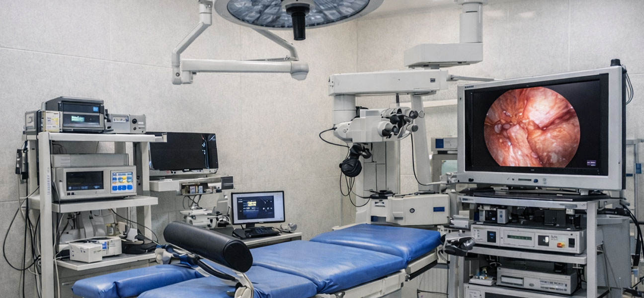
When choosing a reproductive center, patients often focus on the doctor's reputation while overlooking the stability of the laboratory. However, half the success of PGT lies in the lab. The following details are often neglected:
- Blastocyst Culture Rate: Some centers consistently maintain a blastocyst formation rate above 50%, while others achieve only 30%. The difference stems from the culture system, humidity, and operational experience.
- Biopsy Timing and Method: Biopsy on day 5 or day 6, laser aperture size, and the number of cells removed can all affect the embryo's subsequent developmental potential.
- Genetic Counseling Team: The interpretation of mosaicism ratios and segmental deletions in PGT reports requires professional expertise. Centers without genetic counselors may lead to patient misunderstanding of the report.
- Freeze-Thaw Survival Rate: Excellent vitrification techniques can achieve survival rates above 98%, but differences of 2%-5% may exist between laboratories.
Individually, these details seem minor, but cumulatively, they can cause a difference of 10-15 percentage points in the live birth rate per single transfer. It is recommended that patients directly ask the center about its blastocyst formation rate and PGT normal embryo transfer live birth rate for the past year.
6. Common Pitfalls: Misattribution of Success Rates
In outpatient and online consultations, the following misconceptions frequently arise:
- "Someone else succeeded with third-generation IVF, so I will too." — Success rates are population statistics; individual outcomes depend on one's own conditions. Data from peers of the same age is valuable for reference but cannot be directly applied.
- "Third-generation IVF has a higher success rate than first or second generation." — For populations with clear indications, PGT can improve single-transfer efficiency; for those without indications, PGT does not increase the live birth rate and may even harm embryos due to biopsy.
- "As long as I have an embryo, PGT guarantees no miscarriage." — PGT screens for chromosomal aneuploidy but cannot rule out new mutations in single-gene disorders, undetected mosaicism, or miscarriages caused by uterine factors. There is still a 5%-8% risk of miscarriage after PGT.
- "Low AMH means I cannot do third-generation IVF." — Low AMH does not mean it's impossible, but it requires a more refined stimulation protocol and expectation management. AMH of 0.6-1.0 ng/mL may still yield 1-2 blastocysts, but one must be prepared for the possibility of having no normal embryos.
7. Interpretation of Test Indicators: Which Values Directly Correlate with Success Rates?
Before starting a PGT cycle, doctors assess the potential for success using the following indicators:
| Indicator | Reference Range | Impact on Success Rate |
|---|---|---|
| AMH | ≥1.2 ng/mL | Adequate oocyte yield, higher cumulative live birth rate |
| Antral Follicle Count (AFC) | ≥6 | Works synergistically with AMH to assess ovarian reserve |
| FSH | <10 IU/L | Better ovarian response |
| LH | 2-8 IU/L | Abnormal baseline values may indicate PCOS or diminished ovarian reserve |
| Sperm DNA Fragmentation Index (DFI) | <25% | High DFI may lead to decreased blastocyst formation rate |
| Chromosome Karyotype | Normal karyotype | Abnormal karyotypes (e.g., Robertsonian translocation) require PGT-SR |
It is important to note that these indicators are population-based probability references, not absolute thresholds. For example, an AMH of 0.8 ng/mL may still yield normal embryos, albeit with a lower probability. The doctor will make a comprehensive judgment based on age and previous cycle history.
8. Frequently Asked Questions: Common Extended Questions from Patients
The following questions appear most frequently in clinical and online consultations, with unified responses provided below:
8.1 Can third-generation IVF be used for sex selection?
In mainland China, sex selection for non-medical reasons is strictly prohibited. PGT technology can detect embryo sex, but it is only used to avoid sex-linked genetic diseases. Any PGT performed for the purpose of "sex selection" violates regulations.
8.2 If I have third-generation IVF, will my child definitely be healthy?
PGT can screen for common chromosomal aneuploidies, some structural abnormalities, and known single-gene disorders, but it cannot rule out all genetic diseases, new mutations, or birth defects caused by the intrauterine environment. Prenatal diagnosis (amniocentesis) is still necessary after a PGT pregnancy.
8.3 Does PGT embryo biopsy harm the embryo?
Current data indicate that blastocyst biopsy on day 5/6 (approximately 5-8 cells) performed by an experienced operator has minimal impact on the embryo's implantation potential. However, biopsied embryos need to be frozen, and the freeze-thaw process itself carries a very small risk of loss (<2%).
8.4 How long does third-generation IVF take?
A complete PGT cycle typically takes 3-4 months, including: ovarian stimulation (about 12 days), egg retrieval and fertilization, blastocyst culture (5-6 days), biopsy and testing (7-14 days for results), embryo freezing, and scheduled transfer. If using frozen embryos, the fastest time from start to transfer is 2.5 months, but most people require about 3 months due to waiting for reports and endometrial preparation.
8.5 What if there are no normal embryos?
This is the most difficult situation in a PGT cycle. The doctor will analyze the reasons for failure: whether it was due to too few embryos obtained, a low blastocyst formation rate, or an excessively high rate of chromosomal abnormalities. Subsequent options include: adjusting the stimulation protocol for a new cycle, using donor eggs, or abandoning PGT and directly transferring untested embryos (with full informed consent).
Doctor's Advice: How to Rationally View the Success Rate of Third-Generation IVF
As a reproductive specialist, my advice to patients is always: Treat the success rate as a reference line, not the finish line. Before undergoing PGT, first clarify your medical indications, ovarian reserve, and risk tolerance. Consider the following four questions before consulting a doctor:
- What problem am I trying to solve with PGT? (Recurrent miscarriage? Chromosomal abnormality? Single-gene disorder?)
- If I don't obtain a normal embryo during the cycle, can I accept that? Do I have a backup plan?
- Does the reproductive center I choose have an independent genetic counseling team? Are their PGT data from the past year publicly available?
- Does my partner understand the limitations and costs of PGT? Are we in agreement?
Third-generation IVF is a mature embryo screening technology, but it does not promise results. Its true value lies in making every transfer more targeted and helping patients with repeated failures avoid detours. Ultimate success comes from the alignment of medicine, conditions, and decision-making.
Risk Reminder: PGT cannot detect 100% of all chromosomal abnormalities. There is a very low probability (<1%) of misdiagnosis or missed detection of mosaicism. All PGT pregnancies are recommended to undergo prenatal diagnosis (amniocentesis or chorionic villus sampling) in the second trimester.
Examination Reminder: Before starting a PGT cycle, it is essential to complete chromosome karyotyping for both partners, carrier screening for common genetic diseases such as thalassemia and deafness, and an assessment of the uterine environment (hysteroscopy or sonohysterography) to avoid cycle inefficiency due to missed basic examinations.


Comments (0)