

===== Hemophilia Genetic Mechanism and IVF Technology Pathway =====
Hemophilia IVF in China: Genetic Blocking and Preimplantation Genetic Testing Analysis
The core pathway for hemophilia IVF in China is through preimplantation genetic testing—PGT-M technology—to screen for healthy embryos that do not carry the pathogenic gene before embryo transfer. This technology has been maturely applied in China for over fifteen years, and for families of hemophilia carriers with a clearly identified pathogenic gene locus, it can achieve precise genetic blocking.
Hemophilia is divided into Hemophilia A (deficiency of coagulation factor VIII) and Hemophilia B (deficiency of coagulation factor IX), both of which are X-linked recessive genetic diseases. Males have only one X chromosome, and carrying the pathogenic gene leads to the disease; females have two X chromosomes, and carrying the pathogenic gene on one chromosome usually does not cause the disease, but they are carriers. Through PGT-M technology, it is possible to distinguish whether an embryo carries the pathogenic gene and select embryos that do not carry it for transfer, thereby blocking the genetic chain.
===== Why Hemophilia Can Be Genetically Blocked Through IVF =====
Why Hemophilia Can Be Genetically Blocked Through IVF
The inheritance pattern of hemophilia makes it highly suitable for blocking through embryo screening:
- Male patient + Normal female: All sons are normal, and all daughters are carriers (carry the pathogenic gene but do not develop the disease).
- Female carrier + Normal male: Sons have a 50% chance of being hemophilia patients, and daughters have a 50% chance of being carriers.
- Male patient + Female carrier: The inheritance situation is more complex, and the risk of disease in offspring is significantly increased.
The core value of PGT-M technology lies in: before embryo implantation into the uterus, through genetic testing of trophectoderm cells from the blastocyst, the carrier status of each embryo for the hemophilia pathogenic gene is clarified, allowing selection of embryos that do not carry the pathogenic gene for transfer. This is not a treatment for hemophilia, but rather a way to block the transmission of the disease to the next generation at the reproductive genetic level.
Key Prerequisite: The pathogenic mutation site in the family must first be identified through genetic testing, and family verification must be completed. PGT-M is a targeted test for known pathogenic sites and cannot screen for unknown mutations.
===== Doctor's Perspective: Applicable Conditions and Decision Logic of PGT-M Technology =====
Doctor's Perspective: Applicable Conditions and Decision Logic of PGT-M Technology
From a clinical reproductive genetics perspective, families with hemophilia need to meet the following conditions to block inheritance through IVF:
- Clear pathogenic gene locus: The specific pathogenic mutation site on the F8 or F9 gene must first be identified through genetic testing of the proband (patient).
- Complete family verification: Genetic verification of the proband and their parents is required to confirm the inheritance pattern and mutation source. In some cases, this may need to be extended to other family members.
- Adequate ovarian function in the female partner: PGT-M requires obtaining a sufficient number of blastocysts for biopsy. It is generally recommended to have at least 3 to 5 blastocysts available for testing, which imposes certain requirements on the female partner's ovarian reserve.
- Reproductive center with PGT qualification: In China, only medical institutions with the准入资质 (access qualification) for preimplantation genetic testing technology can perform PGT-M. When choosing, the center's qualifications must be verified.
Doctors will emphasize during evaluation: PGT-M is not a treatment for hemophilia, but a method to avoid inheritance by screening embryos. For families with a clearly identified pathogenic gene, the detection accuracy of a single PGT-M cycle for the known site can reach over 95%. However, the overall pregnancy outcome still depends on common factors such as the female partner's age and embryo quality.
===== Actual Process of Hemophilia IVF =====
Actual Process of Hemophilia IVF
Phase 1: Genetic Counseling and Family Verification (1-2 months)
- The proband (hemophilia patient) undergoes coagulation factor activity testing and gene sequencing to identify the pathogenic mutation site.
- Parental verification to confirm the genetic source (de novo mutation or familial inheritance).
- A genetic counseling physician assesses the genetic risk and determines the feasibility of PGT-M and the testing strategy.
Phase 2: Fertility Assessment for Both Partners (2-4 weeks)
- Female partner: AMH, basal FSH, antral follicle count (AFC), chromosome karyotype, infectious disease screening, blood type, liver and kidney function, etc.
- Male partner: Semen analysis, chromosome karyotype, infectious disease screening, etc.
- The assessment results directly determine the ovarian stimulation protocol and the expected number of retrieved oocytes.
Phase 3: Ovarian Stimulation and In Vitro Fertilization (2-3 weeks)
- An individualized ovarian stimulation protocol (antagonist protocol, long protocol, PPOS protocol, etc.) is developed based on the female partner's ovarian function.
- Follicle development is monitored via ultrasound, and HCG is administered at the appropriate time to trigger ovulation.
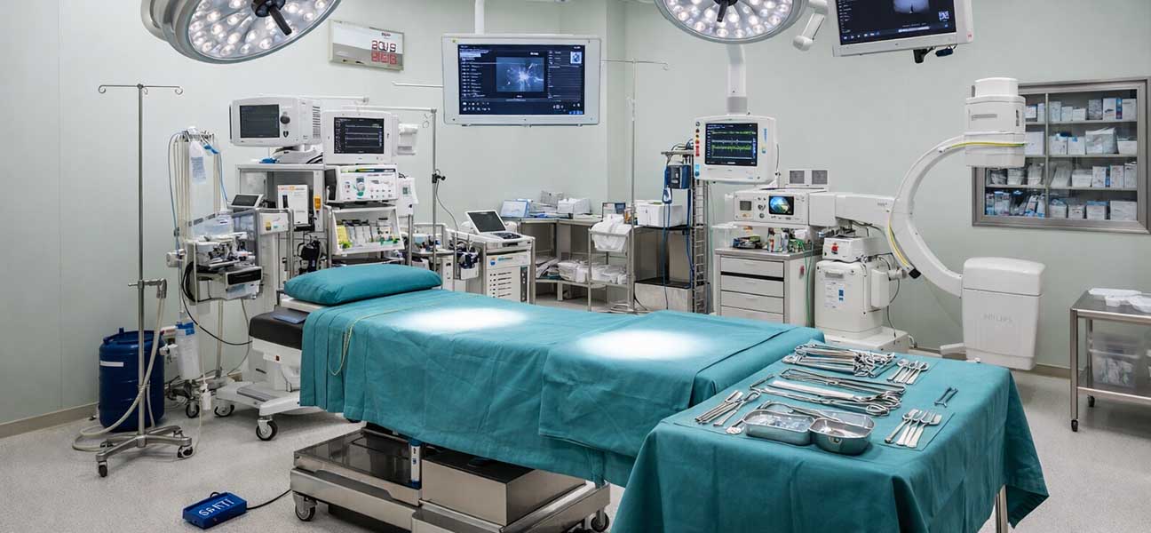
- Egg retrieval surgery (performed under intravenous anesthesia; for hemophilia patients, coagulation management must be coordinated with the hematology department).
- ICSI (Intracytoplasmic Sperm Injection) is used for fertilization to avoid uncertainty regarding the sperm carrying the pathogenic gene.
Phase 4: Embryo Culture and Biopsy (5-6 days)
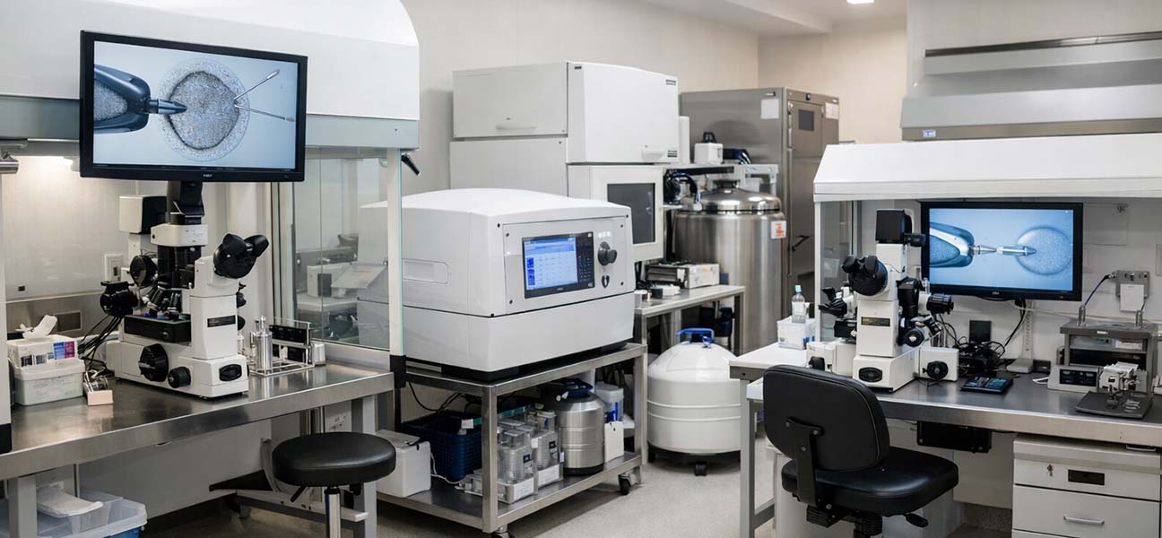
- Fertilized eggs are cultured in the laboratory to the blastocyst stage (day 5-6).
- 5-10 cells are biopsied from the trophectoderm of the blastocyst and sent for genetic testing.
- After biopsy, the embryos are immediately cryopreserved (vitrification) while awaiting test results.
Phase 5: PGT-M Testing (2-4 weeks)
- Whole genome amplification is performed on the biopsied cells to obtain sufficient DNA.
- PCR or NGS methods are used to detect the known pathogenic site, along with linkage analysis for verification.
- An embryo genetic testing report is issued, clarifying the pathogenic gene carrier status of each embryo.
Phase 6: Frozen Embryo Transfer and Prenatal Diagnosis (1-2 months)
- A healthy embryo not carrying the pathogenic gene is selected, thawed, and transferred after the female partner's endometrium is adequately prepared.
- Pregnancy test is performed 12-14 days after transfer.
- After confirming pregnancy, amniocentesis is performed between 16-20 weeks of gestation for prenatal diagnosis to ultimately confirm the fetal genotype.
===== Timeline and Cycle Planning =====
Timeline and Cycle Planning
| Phase | Estimated Time | Key Considerations |
|---|---|---|
| Genetic Counseling & Family Verification | 1-2 months | Requires blood samples from proband and parents; verification cycle may be longer for de novo mutations |
| Fertility Assessment for Both Partners | 2-4 weeks | Some test results have validity periods (e.g., infectious disease screening valid for 6 months) |
| Ovarian Stimulation & Egg Retrieval | 2-3 weeks | Requires coordination with the female partner's menstrual cycle; hemophilia patients need preoperative evaluation by hematology |
| Embryo Culture & Biopsy | 5-6 days | Requires obtaining a sufficient number of blastocysts; cleavage-stage embryos can also be biopsied, but blastocysts are preferred |
| PGT-M Testing | 2-4 weeks | Testing time varies by method and testing center; expedited services can shorten to 10-14 days |
| Frozen Embryo Transfer | 1-2 months | Requires adequate endometrial preparation; natural cycle or hormone replacement cycle are both options |
| Prenatal Diagnosis | 16-20 weeks of gestation | Amniocentesis to ultimately confirm fetal genotype |
The entire cycle, from initiating genetic counseling to completing the transfer, typically takes 3-4 months. If family verification is incomplete, the gene locus is unclear, or repeated verification is needed, the time may extend to 5-6 months.
===== Interpretation of Test Indicators and Clinical Significance =====
Interpretation of Test Indicators and Clinical Significance
Key Test Indicators for Female Partner
- AMH (Anti-Müllerian Hormone): Reflects ovarian reserve; >1.0 ng/mL is normal, <0.5 ng/mL indicates significantly diminished reserve.
- Basal FSH (Follicle-Stimulating Hormone): Measured on day 2-3 of the menstrual cycle; <10 IU/L indicates normal ovarian function, >12 IU/L warrants concern for diminished reserve.
- Antral Follicle Count (AFC): Total antral follicles in both ovaries >5 is normal, <3 suggests potentially poor ovarian response.
- Chromosome Karyotype: To rule out chromosomal structural abnormalities and ensure a clear genetic background for the embryo.
Key Test Indicators for Male Partner
- Semen Analysis: Sperm concentration ≥15×10⁶/mL, progressive motility ≥32%, normal morphology ≥4% (strict criteria).
- Sperm DNA Fragmentation Index (DFI): <30% is normal; high DFI may affect embryo development and pregnancy outcomes.
Key Genetic Indicators
- Coagulation Factor VIII Activity: Hemophilia A patients <40% (severe <1%).
- Coagulation Factor IX Activity: Hemophilia B patients <40% (severe <1%).
- Genetic Testing Report: Identifies the mutation site on the F8 or F9 gene, including mutation type (point mutation, deletion, insertion, rearrangement, etc.) and inheritance pattern.
Note: Coagulation factor activity levels do not affect the IVF procedure itself, but during egg retrieval and transfer, coordination with the hematology department is necessary to manage perioperative coagulation risk.
===== Special Situations Management =====
Special Situations Management
Situation 1: Male Partner is a Hemophilia Patient, Female Partner is Normal
All sons are normal, and all daughters are carriers. PGT-M can select male embryos for transfer to avoid having carrier daughters; or directly screen for embryos that do not carry the pathogenic gene (regardless of sex) to achieve complete blocking.
Situation 2: Female Partner is a Hemophilia Carrier, Male Partner is Normal
Sons have a 50% chance of being patients, and daughters have a 50% chance of being carriers. PGT-M can select embryos that do not carry the pathogenic gene for transfer, regardless of sex. This is the most common clinical scenario.
Situation 3: Male Partner is a Patient and Female Partner is a Carrier
The genetic risk is higher: daughters may be patients or carriers, and sons may be patients or normal. It is essential to screen for embryos that do not carry any pathogenic gene through PGT-M, and it is recommended to combine this with chromosome karyotype analysis.
Situation 4: De Novo Mutation or No Proband Sample Available
If a blood sample from the proband (patient) cannot be obtained, family verification becomes very difficult. Embryo linkage analysis can be used for indirect inference, but accuracy decreases. In such cases, it is recommended to thoroughly discuss the feasibility and limitations of PGT-M with a genetic counseling physician.
Situation 5: Hemophilia Combined with Other Infertility Factors
If there are concurrent factors such as tubal issues, male oligoasthenospermia, or endometriosis, PGT-M needs to be integrated with conventional infertility treatment. The overall plan should be led by reproductive medicine, with genetic counseling support.
===== Frequently Asked Questions =====
Frequently Asked Questions
Q1: What is the success rate of hemophilia IVF?
The detection accuracy of PGT-M technology itself for known pathogenic sites exceeds 95%. However, the overall pregnancy rate is directly related to the female partner's age and embryo quality. The live birth rate per single frozen embryo transfer for women under 35 is about 40%-50%, dropping significantly to 15%-25% for those over 40. The number and quality of embryos are key influencing factors.
Q2: How many embryos are needed for PGT-M?
Clinically, it is recommended to have at least 3-5 blastocysts available for biopsy. If the number of obtained embryos is low (e.g., only 1-2 blastocysts), the possibility of proceeding with PGT-M can be discussed with the doctor, weighing the value of testing against the risk of embryo loss.
Q3: How long does PGT-M testing take?
Routinely, it takes 2-4 weeks. Some reproductive centers offer expedited services in collaboration with genetic laboratories, which can shorten the time to 10-14 days. Embryos must be cryopreserved while awaiting results, so the testing time directly affects the transfer cycle schedule.
Q4: What is the approximate cost of PGT-M for hemophilia?
In China, the total cost for a complete cycle including ovarian stimulation, egg retrieval, ICSI, blastocyst culture, biopsy, PGT-M testing, and one frozen embryo transfer is approximately 50,000 to 80,000 RMB. The exact cost varies by region, hospital level, and individualized medication protocols. Additional cycles or multiple transfers are calculated separately.
Q5: Is prenatal diagnosis still needed after transfer?
Yes. PGT-M is a preimplantation screening and cannot replace prenatal diagnosis. After confirming pregnancy, it is recommended to undergo prenatal genetic diagnosis via amniocentesis between 16-20 weeks of gestation to ultimately confirm the fetal genotype. This is the final verification step to ensure the effectiveness of genetic blocking.
Q6: Is there a risk of bleeding during egg retrieval or transfer for hemophilia patients?
Egg retrieval is an invasive procedure. Hemophilia patients require preoperative evaluation of coagulation status by the hematology department and, if necessary, supplementation of coagulation factors. Transfer is a non-invasive procedure with a very low risk of bleeding. The entire IVF cycle requires joint management by reproductive medicine and hematology.
===== Doctor's Advice =====
Doctor's Advice
For families considering blocking hemophilia inheritance through IVF, it is recommended to proceed step by step as follows:
1. First, complete hemophilia genetic testing at the hematology department of a local tertiary hospital to identify the pathogenic site. Coagulation factor activity testing and gene sequencing are fundamental.
2. Bring the complete genetic testing report and family information to a reproductive center with PGT qualification for genetic counseling. Simultaneously, assess the female partner's ovarian function and fertility status.
3. Work with the reproductive doctor and genetic counselor to develop an individualized PGT-M plan, including the ovarian stimulation strategy, testing platform, and transfer plan.
4. Proceed with the cycle as planned, maintaining communication with the hematology department to ensure proper coagulation management.
5. After transfer, be sure to complete prenatal diagnosis to confirm the fetal genotype. PGT-M is a highly effective screening tool, but it is not a 100% absolute diagnosis.
Special Reminder: During the IVF cycle, families with hemophilia must pay close attention to the patient's coagulation function management. Before egg retrieval surgery, a perioperative coagulation factor supplementation plan must be developed in collaboration with the hematology department. PGT-M technology cannot correct the patient's own hemophilia; it only acts on embryo screening. The patient's own coagulation disorder still requires lifelong standard treatment.







Comments (0)