

AI Summary
Main Content Begins
Reproductive Medicine Knowledge Editor · Clinical Observation Notes
Real Consultation Scenarios in Outpatient Clinics
"Doctor, I heard there is an incubator called EmbryoScope that can continuously record videos of embryo development. Is this technology mature in China? Do hospitals here have it? I've had two failed transfers. Would this be helpful?" — This is an increasingly common question in reproductive medicine clinics. With the spread of information about assisted reproductive technology among patients, time-lapse incubators are gradually entering the public eye. However, there is still significant information asymmetry regarding what this technology solves, who it is suitable for, and its current implementation in China.
What is EmbryoScope Technology
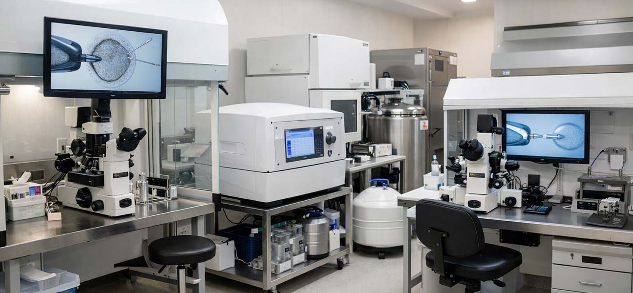
EmbryoScope is a time-lapse imaging incubator developed by a Danish company (UnisenseFertiliTech, now part of the Vitrolife Group). It integrates a micro-camera system and LED light source into a standard incubator, automatically capturing high-resolution images every 5–10 minutes during normal embryo culture and continuously recording the entire development process (typically 5–7 days until the blastocyst stage).
The key difference from traditional incubators is that traditional methods require embryologists to remove embryos from the incubator at fixed time points (e.g., day 1, 2, 3, and 5 post-fertilization) for static morphological assessment under a microscope. In contrast, EmbryoScope obtains complete developmental timing information without removing the embryos.
Current Development Status in China
As of 2024, more than 40 reproductive medicine centers in China have introduced time-lapse imaging incubators, with the EmbryoScope series holding a major share. These centers are mainly distributed in the following regions:
- Large public hospitals in first-tier cities (Well-known reproductive centers in Beijing, Shanghai, Guangzhou, Shenzhen)
- Provincial key assisted reproduction laboratories (Large reproductive centers in Hangzhou, Chengdu, Wuhan, Nanjing, Jinan, etc.)
- Some high-end private fertility clinics (Primarily serving complex cases and advanced-age populations)
In terms of application depth, about 60% of the centers that have introduced the technology use it for patients with specific indications (e.g., recurrent implantation failure, slow embryo development, advanced maternal age, previous cycles with no available embryos for transfer). About 30% of centers use it as a routine culture protocol in some cycles, while the remaining 10% are still in the technology validation and personnel training phase.
Technical Advantages and Clinical Value
Additional Information Provided by Dynamic Assessment
Traditional static assessment only provides a "snapshot" of the embryo at a specific time point, whereas time-lapse imaging can observe:
- Fertilization time (IP): Time from insemination to pronuclei appearance. Abnormal delays may indicate fertilization abnormalities.
- First cleavage time (t2): Common window is 25–27 hours. Too early or too late is associated with reduced developmental potential.
- Second and third cleavage times (t4, t8): Regular cleavage is an important marker of embryo health.
- Morula formation time (tm): Typically 80–90 hours post-fertilization.
- Blastocyst formation time (tB): Common range is 100–120 hours; expansion speed is also evaluated.
- Abnormal events such as reverse cleavage and direct cleavage: These are easily missed in static assessment.
These dynamic parameters can help embryologists more accurately select embryos with high implantation potential, especially when morphological scores are similar. The additional information provided by time-lapse imaging offers distinguishing value.
Overview of Clinical Research Evidence
Multiple published observational studies and meta-analyses show that compared to conventional culture, using time-lapse imaging for embryo selection leads to a mild to moderate improvement in implantation and clinical pregnancy rates, along with a reduction in cycle cancellation rates. However, it should be noted that large-scale, multi-center, randomized controlled trials confirming a significant advantage in live birth rates are still lacking. The evidence for benefit in patients with recurrent implantation failure is relatively more robust.
Suitable and Unsuitable Populations
| Suitable Situations | Unsuitable or Unnecessary Situations |
|---|---|
| Recurrent implantation failure (≥2 failed transfers) | Very few embryos (only 1–2), limited statistical value of dynamic assessment |
| Previous abnormal embryo development (slow cleavage, high fragmentation, low blastocyst formation rate) | Limited financial resources, and conventional morphological assessment can already identify clearly good-quality embryos |
| Advanced maternal age (≥38 years) with a larger number of embryos requiring fine selection | PGT-A (Preimplantation Genetic Testing for Aneuploidy) already decided, genetic screening is the primary criterion |
| Research or quality control scenarios requiring precise assessment of embryo developmental timing | Laboratory lacks trained embryologists for data interpretation |
| Cycles requiring extremely high stability of the culture environment | Patient does not accept any additional technical intervention |
Comparison with Traditional Incubators
| Comparison Dimension | Traditional Incubator | EmbryoScope Time-Lapse Imaging Incubator |
|---|---|---|
| Assessment Method | Remove embryos at fixed time points for static observation | Continuous imaging inside the incubator, dynamic recording |
| Culture Environment Disturbance | Each opening causes brief fluctuations in temperature and gas concentration | No need to open the incubator, higher environmental stability |
| Data Volume | 3–5 time point data per embryo | 1000–2000 images per embryo |
| Equipment Cost | Relatively low | 2–3 times higher than conventional incubators |
| Personnel Training Requirements | Standard embryology training sufficient | Additional training required for time-lapse imaging analysis and parameter interpretation |
| Target Population | Basic protocol for all IVF cycles | Priority for complex cases; some centers use for all cycles |
Actual Usage Process
A complete EmbryoScope usage cycle includes the following steps:
- Preparation Phase: Pre-equilibration of culture media (at least 4 hours), labeling dedicated culture dishes (usually 12-well or 6-well plates) with patient information, and preheating in the incubator.
- Embryo Loading: After egg retrieval, perform conventional IVF or ICSI with mature oocytes and sperm, then transfer embryos or zygotes to the dedicated culture dish, placing one embryo per well and covering with an appropriate amount of culture oil.
- Data Acquisition: Close the incubator door and start the imaging program. The incubator automatically controls temperature (37.0±0.2°C), CO₂ concentration (6.0%), and O₂ concentration (5.0%), capturing images every 5 minutes.
- Data Analysis: Embryologists review the video at fixed time points (or daily) during the culture period using the accompanying software, marking key developmental events. The system automatically calculates time parameters.
- Embryo Assessment and Selection: Combine morphological scores and time-lapse parameters with clinical information such as patient age and history to select embryos for transfer or blastocysts suitable for freezing.
- Transfer or Freezing: Perform embryo transfer on day 3 or day 5–6, or vitrification for cryopreservation.
The entire process requires high standards of laboratory workflow management, especially regarding data recording norms and consistency in embryologist interpretation.
Interpretation of Key Evaluation Parameters
The main parameters provided by time-lapse imaging technology and their clinical significance are as follows:
| Parameter Abbreviation | Definition | Common Normal Range | Abnormal Indication |
|---|---|---|---|
| IP | Fertilization time (insemination to pronuclei appearance) | 20–24 hours | >26 hours may indicate fertilization abnormality or sperm factor |
| t2 | First cleavage time | 25–27 hours | <22 hours or >30 hours associated with low implantation rate |
| t4 | Second cleavage (2→4 cells) | 36–40 hours | Delay may indicate cytoskeletal or chromosomal abnormalities |
| t8 | Third cleavage (4→8 cells) | 48–54 hours | Irregular cleavage or direct cleavage (1→3) requires caution |
| tm | Morula formation time | 80–90 hours | >96 hours indicates reduced developmental potential |
| tB | Blastocyst formation time | 100–120 hours | >125 hours, blastocyst quality is usually poor |
| tEB | Blastocyst expansion time | 110–130 hours | Slow expansion is related to inner cell mass quality |
It must be emphasized that the interpretation of these parameters must be combined with the overall embryo morphology, patient age, cause of infertility, and other factors; they cannot be used in isolation. Different embryo culture systems and laboratory environments may cause shifts in parameter ranges, and each center should establish its own reference data.
Most Common Information Misconceptions
- Misconception 1: "Using EmbryoScope guarantees success" — Time-lapse imaging optimizes selection accuracy, does not change embryo quality, and is ineffective for implantation failure caused by chromosomal abnormalities.
- Misconception 2: "All hospitals have it, and it's the same everywhere" — Reproductive centers in China with EmbryoScope are still a minority, and there are differences in equipment models, software versions, and personnel experience among centers.
- Misconception 3: "Time-lapse imaging can replace embryologists" — On the contrary, time-lapse imaging generates far more data than traditional methods, requiring higher analytical skills from embryologists.
- Misconception 4: "It must cost extra, so it must be better" — Some centers charge an additional fee for time-lapse imaging (ranging from approximately 3000–8000 RMB), but the fee level does not represent the technology's effectiveness. Patients should understand the specific evaluation plan.
Observations from Practitioners
From the perspective of the embryology laboratory, time-lapse imaging technology has indeed changed workflows and thought processes. In traditional methods, embryologists make "instant judgments" at fixed time points, whereas time-lapse imaging forces observers to understand the continuity of development — the change in an embryo between 30 and 32 hours may determine its suitability for transfer. This temporal dimension moves embryo assessment from "scoring" to "understanding the developmental trajectory."
However, it must also be acknowledged that time-lapse imaging is not without limitations. First, the high equipment and maintenance costs make it difficult for some centers to adopt widely. Second, data standardization is an issue: parameter definitions and algorithms vary between different brands and software, making direct comparison of data between centers difficult. Third, for cycles with very few embryos (e.g., only 1–2 oocytes retrieved), the statistical advantage of time-lapse imaging is difficult to realize.
In actual clinical practice, the most stable value of time-lapse imaging lies in "exclusion" rather than "selection" — that is, using abnormal developmental parameters to exclude embryos that appear morphologically normal but have abnormal developmental trajectories. This is particularly prominent in patients with recurrent implantation failure.
Frequently Asked Questions
Q: Which hospitals in China have EmbryoScope?
A: They are mainly distributed in large provincial reproductive centers and some high-end private clinics, such as Peking Union Medical College Hospital, Peking University Third Hospital, Renji Hospital Affiliated to Shanghai Jiao Tong University School of Medicine, CITIC-Xiangya Reproductive and Genetic Hospital, and the Women's Hospital School of Medicine Zhejiang University, among more than 40 institutions. It is recommended to call the target hospital's reproductive center to confirm specific availability.
Q: What additional preparations are needed for using EmbryoScope?
A: No additional preparation is needed for patients. The egg retrieval, fertilization, and culture processes are the same as conventional IVF. The difference is that embryologists use dedicated culture dishes and incorporate time-lapse parameters in the assessment.
Q: What is the difference in pregnancy rates between EmbryoScope and conventional incubators?
A: Existing studies suggest that in patients with recurrent implantation failure, the implantation rate in the time-lapse imaging group may increase by about 8–15 percentage points. However, the difference is smaller in the general IVF population, around 3–8 percentage points. The specific effect is highly influenced by patient selection and laboratory standards.
Q: Is it safer to culture embryos in EmbryoScope?
A: From the perspective of environmental stability, reducing the number of times the incubator is opened helps maintain stable culture conditions. However, safety depends not only on the type of incubator but also on the overall quality control system of the laboratory, including air quality, culture media quality control, and aseptic techniques.
Editor's Note: This article is based on publicly available clinical data and industry observations as of the end of 2024 and does not constitute specific medical advice. Decisions regarding the application of time-lapse imaging technology should be made based on the patient's individual circumstances, the center's equipment conditions, and the physician's professional judgment. Technical parameters and clinical evidence may be updated with research progress. It is recommended to regularly consult the latest literature or reproductive medicine professionals.



Comments (0)