

Author: Reproductive Specialist | Source: Assisted Reproduction Knowledge Base | Reading Time: Approx. 8 minutes
A 36-year-old woman came to the clinic with a history of four first-trimester miscarriages. She had undergone tests for sex hormone six items, thyroid function, blocking antibodies, and chromosome karyotype analysis for both partners, all of which were reported normal. She asked several direct questions: "Can going abroad for IVF solve my problem? Can PGT prevent miscarriage? Which hospital should I choose?" Behind these questions lies uncertainty about the root cause of recurrent miscarriage and confusion due to the information asymmetry in overseas medical care.
Why Systematic Screening for the Causes of Recurrent Miscarriage is Necessary
Recurrent pregnancy loss (RPL) occurs in about 1%–3% of the reproductive-age population, defined as two or more pregnancy losses before 20 weeks of gestation. The etiology is complex, and no single test can cover all possibilities. Clinical statistics show that among RPL cases with a clear cause, chromosomal abnormalities account for about 30%–40%, uterine anatomical abnormalities for 10%–15%, immune factors for 20%–30%, coagulation abnormalities for 10%–15%, and endocrine and infectious factors each account for 5%–10%. In about 30%–40% of cases, the cause remains unidentified.
Different etiologies require completely different intervention paths. Chromosomal abnormalities primarily rely on PGT technology to screen for normal embryos; uterine anatomical abnormalities require hysteroscopic surgical correction; immune factors need immunomodulatory therapy; coagulation abnormalities require anticoagulation regimens. Therefore, the first criterion for choosing an overseas IVF hospital is not the success rate number, but whether the hospital has a complete RPL etiological screening capability and multidisciplinary collaboration.
Core Criteria for Choosing an Overseas IVF Hospital
For patients with recurrent miscarriage, the selection of an overseas IVF hospital should be evaluated based on the following four dimensions:
- Completeness of Etiological Screening: Does the hospital routinely perform chromosome karyotype analysis for both partners, hysteroscopy, antiphospholipid antibody panel, protein C/S activity, thyroid function and autoantibodies, and chronic endometritis (CD138+) testing? Missing any one item could lead to missed diagnosis of the cause.
- Accessibility of PGT Technology: Does the hospital have the capability for PGT-A (aneuploidy screening), PGT-SR (structural rearrangement screening), and PGT-M (monogenic disease screening)? For recurrent miscarriage, PGT-A is the most basic requirement, but it is important to note that PGT cannot solve immune or coagulation issues.
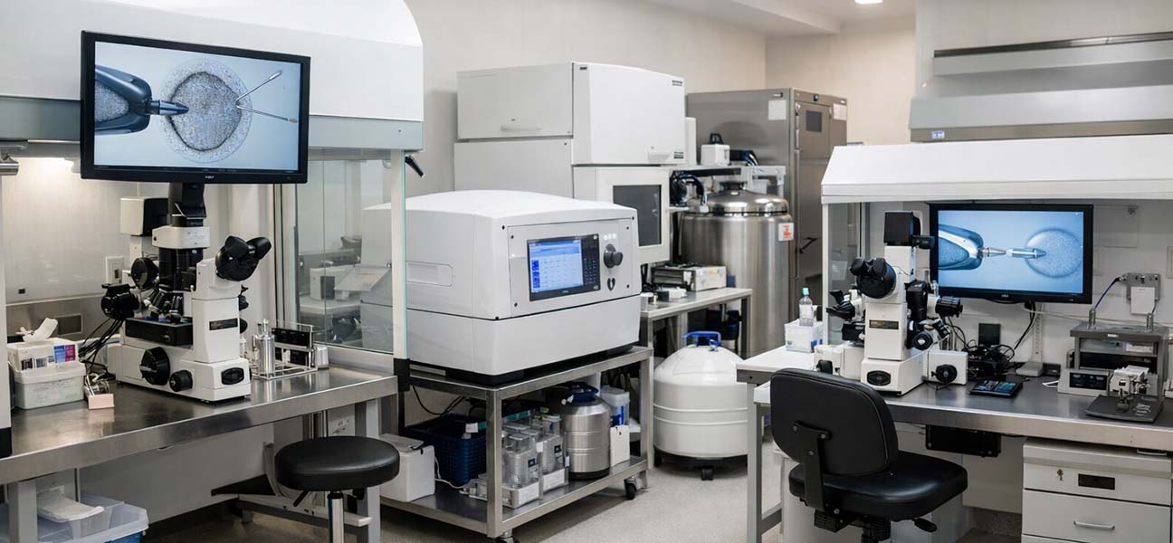
- Laboratory Stability: Are the embryology lab's fertilization rate, blastocyst formation rate, and survival rate after thawing publicly available? Laboratory conditions directly affect embryo developmental potential and the accuracy of PGT results.
- Multidisciplinary Collaboration Mechanism: Does the hospital have weekly joint discussions involving reproductive medicine, genetic counseling, immunology, hematology, and hysteroscopy specialists? RPL patients often require a multidisciplinary pre-transplant preparation plan.
Interpretation and Clinical Significance of Key Examination Indicators
Chromosome Karyotype Analysis for Both Partners
Standard karyotype analysis (G-banding) can detect numerical chromosome abnormalities and structural abnormalities larger than 5Mb. If one partner is a carrier of a balanced translocation or Robertsonian translocation, the risk of embryonic chromosomal abnormalities is significantly increased. In this case, PGT-SR is a clear indication. It is important to note that routine karyotype analysis cannot detect microdeletions and microduplications smaller than 5Mb; SNP-array or CMA should be added when necessary.
Uterine Cavity Evaluation
Hysteroscopy is the gold standard, capable of detecting endometrial polyps, intrauterine adhesions, uterine septum, and submucosal fibroids. The detection rate of uterine cavity abnormalities in patients with recurrent miscarriage is about 15%–25%. Additionally, chronic endometritis (CE) can be diagnosed by endometrial biopsy showing CD138+ plasma cell infiltration. CE is associated with both recurrent implantation failure and miscarriage, and outcomes can improve after antibiotic treatment.
Immune Factor Screening
The standard panel should include: anticardiolipin antibodies (IgG/IgM), β2-glycoprotein I antibodies, lupus anticoagulant, antinuclear antibodies, thyroid peroxidase antibodies, and thyroglobulin antibodies. If these indicators are positive, anticoagulation or immunomodulatory therapy should be initiated under the guidance of a rheumatology or immunology specialist. The clinical value of NK cell activity testing and T cell subset analysis remains controversial and is not considered routine.
Coagulation Function Tests
Protein C activity, Protein S activity, Antithrombin III, Factor V Leiden mutation, Prothrombin G20210A mutation, and homocysteine levels. Patients with thrombophilia have an increased risk of thrombosis during pregnancy, and placental microthrombosis can lead to miscarriage. Anticoagulation therapy with low molecular weight heparin or aspirin is required.
Male Factor Assessment
Sperm DNA fragmentation index (DFI > 30% is associated with miscarriage risk), sperm nuclear protein conversion, Y chromosome microdeletion. Elevated DFI may be related to oxidative stress, varicocele, or infection. Lifestyle adjustments or antioxidant therapy 3–6 months before treatment can improve the condition.
| Examination Item | Clinical Significance | Recommended Timing |
|---|---|---|
| Chromosome Karyotype Analysis | Detects structural abnormalities like balanced translocations, Robertsonian translocations | Before attempting pregnancy or after first miscarriage |
| Hysteroscopy | Evaluates uterine cavity shape and endometrial environment | 3–7 days after menstruation ends |
| Antiphospholipid Antibody Panel | Diagnoses Antiphospholipid Syndrome | Non-pregnant state or early pregnancy |
| Protein C/S Activity | Screens for hereditary thrombophilia | Non-pregnant state (after stopping anticoagulants) |
| Sperm DNA Fragmentation Index | Assesses sperm nuclear DNA integrity | After 2–5 days of abstinence |
Details Most Easily Overlooked in Recurrent Miscarriage Evaluation
- Chromosome analysis is needed for both partners, not just the woman. Male carriers of balanced translocations usually have a normal phenotype, but the risk of embryonic chromosomal abnormalities can be as high as 50%–70%.
- Hysteroscopy cannot be replaced by 3D ultrasound. 3D ultrasound can preliminarily assess uterine cavity shape, but it cannot directly observe endometrial inflammation or minor adhesions; hysteroscopy is the gold standard.
- Antiphospholipid antibodies need repeat testing for confirmation. A single positive result does not diagnose APS; a positive result repeated after an interval of at least 12 weeks is clinically significant.
- Culture and biopsy for chronic endometritis require high standards. Routine endometrial biopsy easily misses the diagnosis; CD138 immunohistochemical staining combined with bacterial culture is necessary.
- Control TSH below 2.5 mIU/L. Even if thyroid function is within the normal range, TSH > 2.5 is associated with an increased risk of early miscarriage.
- Normal semen analysis does not guarantee normal chromosomes or DNA integrity. Even with normal sperm morphology and density, there may still be elevated DFI or Y chromosome microdeletions.
Common Pitfalls When Choosing an Overseas IVF Hospital
Mistake 1: Believing PGT-A can solve all miscarriage problems. PGT-A only screens for embryonic chromosomal aneuploidy; it is ineffective for miscarriages caused by immune factors, coagulation abnormalities, or uterine environment issues. Some patients still miscarry after transferring PGT-A normal embryos because non-chromosomal causes were overlooked.
Mistake 2: Only looking at the hospital's published "success rate." Success rates are influenced by multiple factors such as age, egg source, cycle type (fresh/frozen), and transfer strategy, and statistical methods vary between hospitals. For RPL patients, it is more important to check if the hospital has a dedicated RPL diagnostic and treatment pathway and genetic counseling capability.
Mistake 3: Choosing a hospital without complete screening capabilities. Some overseas hospitals market "quick cycle initiation" and do not require patients to complete basic screening. Starting ovarian stimulation without identifying the cause in recurrent miscarriage patients may lead to repeated failed cycles.
Mistake 4: Ignoring visa and document timelines. Overseas IVF involves passport validity (usually >6 months), visa type (medical visa requires a hospital invitation letter), marriage certificate notarization and translation, and some countries require proof of fertility. Document preparation needs to be completed 2–3 months in advance.
Doctor's Decision-Making Logic: The Path from Etiology to Treatment
When facing patients with recurrent miscarriage, clinical decisions follow the principle of "etiology stratification—intervention stratification":
- Confirmed chromosomal abnormality (in partners or embryo): Priority recommendation for PGT-SR or PGT-A, while assessing ovarian reserve (AMH, antral follicle count) to determine the stimulation protocol.
- Confirmed uterine factor: First perform hysteroscopic surgery for correction (polyp removal, adhesiolysis, septum resection). Re-evaluate uterine cavity shape after 3 months post-surgery before considering transfer.
- Confirmed immune or coagulation abnormality: Collaborate with rheumatology/immunology or hematology to develop a pre-treatment plan. Low molecular weight heparin + aspirin is the basic regimen; some patients may require hydroxychloroquine or corticosteroids.
- Unexplained etiology (Idiopathic RPL): May attempt PGT-A combined with frozen embryo transfer, along with endometrial microbiome testing and endometrial receptivity array (ERA) analysis. However, patients should be informed that there is still a 20%–30% risk of miscarriage.
Medical Treatment Process for Recurrent Miscarriage Patients Undergoing Overseas IVF
Phase 1: Remote Pre-screening (1–2 months)
- Submit previous examination reports from both partners (chromosome, hysteroscopy, immune, coagulation, semen analysis, etc.).
- The hospital's reproductive specialist + genetic counselor jointly assess missing items and create a supplementary examination checklist.
- After completing supplementary tests, the case is submitted for multidisciplinary team (MDT) discussion to clarify the preliminary diagnosis and intervention direction.
Phase 2: Document and Travel Preparation (1–2 months)
- Confirm passport validity (recommended remaining validity > 12 months).
- Apply for a medical visa (some countries require the hospital to provide a treatment plan and cost estimate).
- Marriage certificate notarization + translation + dual apostille (depending on the destination country's requirements).
- File creation materials: ID documents for both partners, marriage certificate, previous medical records summary (translated).
Phase 3: Treatment Abroad (4–6 weeks)
- Menstrual cycle day 2–3: Baseline hormones (FSH, LH, E2, P), AMH, antral follicle count, start ovarian stimulation protocol.
- Ovarian stimulation period: 8–12 days, monitor hormones and follicle development every 2–3 days.
- Egg retrieval day: Transvaginal ultrasound-guided egg retrieval, simultaneous sperm collection.
- Embryo culture + PGT: Blastocyst culture for 5–7 days, biopsy of trophectoderm cells for testing, waiting period approximately 4–6 weeks (can wait back home).
- Transfer preparation: Frozen embryo transfer cycle, endometrial preparation for 8–12 days, pregnancy test 12 days after transfer.
| Phase | Time | Key Actions |
|---|---|---|
| Remote Pre-screening | 1–2 months | Complete missing tests, MDT discussion |
| Document Preparation | 1–2 months | Passport, visa, notarization, translation |
| Ovarian Stimulation + Egg Retrieval | 2–3 weeks | Hormone monitoring, follicle tracking |
| Embryo Culture + PGT | 4–6 weeks | Blastocyst culture, genetic testing |
| Frozen Embryo Transfer | 2–3 weeks | Endometrial preparation, transfer, pregnancy test |
Differences in Hospital Selection by Country
| Country/Region | PGT Technology Maturity | Immune Screening Capability | Cost Reference (RMB) | Cycle Waiting Time |
|---|---|---|---|---|
| USA | High (Full PGT-A/SR/M coverage) | Comprehensive multidisciplinary collaboration | 250,000 – 350,000 | Longer (requires scheduling) |
| Thailand | Medium-High (Primarily PGT-A) | Some hospitals have specialized programs | 100,000 – 180,000 | Shorter |
| Japan | Medium (PGT-A available) | Focuses on detailed management | 150,000 – 220,000 | Moderate |
| Malaysia | Medium-High (PGT-M available) | Moderate comprehensive ability | 100,000 – 150,000 | Moderate |
| Georgia | Medium (PGT-A developing) | Primarily basic screening | 80,000 – 120,000 | Shorter |
Risks to Clarify Before Planning
The etiological screening for recurrent miscarriage is a dynamic process. Some patients may need 1–2 cycles to identify the core issue. Overseas medical treatment involves language barriers, differences in medical systems, and cross-disciplinary coordination. It is recommended to have at least one tripartite consultation with a reproductive specialist, genetic counselor, and rheumatologist/immunologist before starting.
Although PGT technology can significantly reduce the risk of miscarriage caused by chromosomal abnormalities, it cannot completely prevent it. Early pregnancy monitoring after transfer is still necessary, including ultrasound to confirm fetal heartbeat at 6–7 weeks, NT screening at 10–12 weeks, and subsequent prenatal diagnosis.
All overseas hospital choices must be based on the individual's specific etiology and physical condition, not on general rankings. Age, ovarian reserve, previous surgical history, and comorbidities all affect the treatment path and prognosis. It is recommended to bring complete medical records for a personalized evaluation.
Knowledge Base Update Note: This article is compiled based on clinical practice in assisted reproduction and domestic/international guidelines for 2024–2025. The content is intended for educational reference only and does not constitute medical advice. Please follow the opinion of your attending physician for specific diagnosis and treatment plans.







Comments (0)